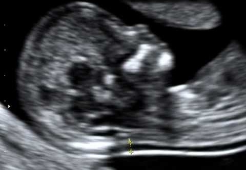
What is Foetal Medicine?
Fetal Medicine primarily deals in providing care to unborn babies. It involves diagnosis and treatment of complications which may arise in unborn babies. As fetal medicine specialists we are specialised in performing:
- Antenatal ultrasound examination of the unborn baby i.e. fetus
- Monitor the proper growth of fetus according to the gestation of the pregnancy
- Look for the normal development of the fetus, detect abnormalities if any
- Guide and provide counselling to the couple undergoing the necessary tests while taking any decisions in case of high risk pregnancies
Fetal Medicine Helpline: +91-9838887030
What we do?
We are specialied in ultrasound examination of the fetus and treatment of complications which may arise in unborn babies.
Service Offered
- Early pregnancy scan
- First trimester screening for chromosomal abnormalities (NT/ NB scan and maternal serum biochemistry) (11 – 13+6 weeks)
- Anomaly scan (TIFFA scan) with screening fetal echo
- Fetal echo
- Growth scan/ Fetal well being scan
- Obstetric Doppler
- Amniocentesis
- Chorionic Villous sampling
- Fetal blood sampling
- NIPT (cf Fetal DNA)
- Genetic Counselling
Fetal Therapy
- Selective fetal reduction
- Interstitial laser/ RFA in complicated twin pregnancies
- Intrauterine Fetal blood transfusions
- Amniodrainage/ Amnioreduction
- Pleurocentesis/ Thoracoamniotic shunt placement
- Vesicoamniotic shunt placement
Early pregnancy scan
This is the first ultrasound examination of the pregnant female. It is done to confirm the location of the pregnancy and to ascertain viability. The fetus is seen for the first time in this scan.
First trimester screening for chromosomal abnormalities

This scan is done between 11 weeks to 13 weeks and 6 days when the fetal length is between 45 – 84 mm. The risk for chromosomal abnormalities in particular Down syndrome (Trisomy 21) is assessed based on the findings of the scan which include the thickness of the skin and the fluid underneath behind the neck (NT), presence or absence of nasal bone, fetal heart rate and other findings. It is then combined with the maternal serum biochemistry to provide improved accuracy for risk assessment.
Other benefits of the 11–13+6 weeks scan include accurate dating of the pregnancy, early diagnosis of major fetal abnormalities, and the detection of multiple pregnancies. The early scan also provides reliable identification of chorionicity, which is the main determinant of outcome in multiple pregnancies.
Anomaly scan
An anomaly scan is detailed study of the structures of the baby. It is done to find out if there is any abnormality in the developing baby and to decide on its cure and precautions throughout the pregnancy and also during child birth. It also helps us assess the risk of chromosomal abnormalities and should be combined with the first trimester screening to improve the accuracy.
Certain abnormalities in the fetus may be associated with chromosomal and genetic abnormalities and have a bearing on the future pregnancies. In case of detection of any structural abnormality in the fetus, further steps can be taken to try and identify the cause and provide counselling about the same.
Fetal echocardiography
A fetal echo or echocardiography is detailed ultrasound study of the structure and the function of the heart of the unborn baby. If any previous ultrasound examinations raised a suspicion of an abnormality in the fetal heart, detailed fetal echocardiography is recommended. If the mother is diabetic, or has taken any medications, or there is family history of cardiac defect then with detailed echo a prenatal diagnosis of fetal congenital heart disease (CHD) shows a significant effect on prenatal and postnatal management of childbirth, care and its outcomes.
Fetal Growth/ Well being
This ultrasound scan is usually carried at about 28-36 weks of pregnancy and aims at determining the growth and health of the fetus.
A pathological decrease in the rate of fetal growth and blood flow defines Fetal Growth Restriction. There may be various causes of this fetal growth restriction which need to be figured out by a fetal medicine expert.
Obstetric Doppler is the study of the blood flows to the fetus.
Amniocentesis
This test is used to diagnose any prenatal chromosomal, genetic abnormalities in the fetus and fetal infections. It is usually performed between 16 and 20 weeks of pregnancy. This test is primarily for those expecting mothers who are at increased risk for genetic and chromosomal problems. The most common reason to have an amniocentesis test is to determine whether a baby has chromosomal abnormality such as Down syndrome.
A needle is passed through the abdominal wall and through the wall of the uterus (womb) into the amniotic fluid under continuous ultrasound guidance. A small amount of fluid is aspirated into the syringe and the needle is withdrawn. The amniotic fluid is then sent for further analysis to the laboratory.
Chorionic Villous sampling
It is a test usually offered during first trimester of pregnancy to detect chromosomal abnormality or genetic syndrome. CVS test is performed by taking a sample of the chorionic villi (placental tissue). The genetic information in the tissue helps in finding out if an abnormal genetic condition is present like Down syndrome, Thalassemia or other genetic syndrome. This helps them to decide how to proceed with the pregnancy.
Fetal Reduction
Being pregnant with two or more babies has its own challenges. These pregnancies are more prone for pre term deliveries and fetal growth restriction. A procedure to lower the number of foetuses and increase the chances of healthy pregnancy and survival is called selective fetal reduction. The more babies a lady carries, the more are the chances of having a miscarriage, preterm delivery or still birth.
Why Choose Apollomedics
Department of Fetal Medicine at Apollomedics hospitals provides comprehensive care to the unborn fetus nourishing inside the pregnant mother to ensure a healthy baby after delivery. We are skilled to diagnose and treat ailments related to the pregnant mother and the fetus at world class diagnostic setup with highly experienced medical professionals.
Frequently Asked Questions
In a normal and uncomplicated pregnancy, the following scans are required:
1). Fetal viability scan
2). First trimester screening (NT/NB scan) at 11-14 weeks
3). TIFFA/ Anomaly scan/ Level II scan at 18-20 weeks
4). Fetal well being scan at 32-34 weeks
Ultrasound scans use sound waves which are absolutely safe for the baby and no detrimental effects of the same have ever been noted.
It is a specialised ultrasound done at 11- 14 weeks to:
1). Check the growth of the fetus
2). Examine the structures of the fetus
3). Calculate the risk of chromosomal abnormalities in the fetus
Apollo lucknow is the best hospitals to perform NT/NB scan in lucknow uttar Pradesh.
It is done in conjunction with The NT/ NB scan to better compute the risks of chromosomal abnormalities. It need mother’s blood sample along with the NT/ NB scan report.
Apollo lucknow is the best place to have double marker test.
It is the detailed ultrasound study of the fetal heart.
Apollo medics is the best hospital to conduct fetal echo in lucknow uttar Pradesh.
It is best performed at 22 – 24 weeks.
TIFFA is a specialised ultrasound scan and needs to study the baby’s structures in detail. It depends on the fetal position and if the baby is not in optimal position for the scan, it might take multiple sittings to complete the scan.
TIFFA is a specialised ultrasound scan and needs to study the baby’s structures in detail. It depends on the fetal position and if the baby is not in optimal position for the scan, it might take multiple sittings to complete the scan.
Apollo medics is the best hospital to conduct TIFFA test in lucknow uttar Pradesh.
Identity proof of the mother (photo and address should be mentioned)
Prescription of the doctor for the scan
The patient needs to hold urine only for the fetal vibility scan i.e the first scan done at 6-8 weeks. Full bladder is not required for any other fetal ultrasound examination
The mother should always eat and come to the hospital. No fetal examinations require the mother to be empty stomach
Doctor’s Profile


